
The impact achieved
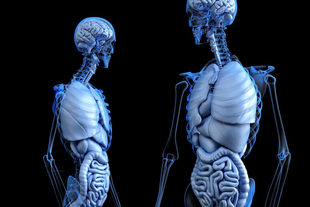
This research project has pioneered the use of Electrical Impedance Tomography (EIT), which can be used to image organ function in real time (100 images/second). Compared with existing technology it is highly portable, inexpensive and lends itself readily to remote imaging to save lives. The project’s key impacts are:
- Provision of imaging algorithms and clinical analysis impacting on clinical software
- Creation of the largest clinical data store for EIT clinical data in the world (> 50TBytes) for use by clinicians and industrial/academic researchers
- Development of new wearable hardware for application on patents impacting on clinical usability of EIT
- Successful use for monitoring pre-term neonates in the largest clinical study undertaken to date and for identifying key parameters for the clinical management of neonates with respiratory conditions impacting on clinical practices
- Cost saving of €928 to €10,705 per patient in the Dutch setting, or €1,124 to €8,496 per patient in the German setting
- Ongoing work with Printed Electronics Limited (PEL), a UK-based technology company, to develop print on flexible printed circuits for the EIT neonate system.
The research behind it
The impact described above evolved from a series of specific developments employing EIT, including:
- Successful generation of the first 2D images of impedance change inside the human head using EIT (1996 –2003)
- A range of clinical applications and the development of a method of automatically generating subject-specific FE models (2003-2007) and, since 2008, the development of a further algorithm
- Development of algorithms and hardware for image reconstruction, parameter measurement and boundary form generation (since 2016), culminating in the first large scale study monitoring the lung function of 200 neonates (preterm, high risk) for 72 hours each
- Clinical system for use in neonatal intensive care units, and further developments to clinical hardware for bedside monitoring of lung gestation of pre-term neonates.
The research continued to flourish and diversify throughout the coronavirus pandemic when we repurposed the hardware and techniques for monitoring COVID-19 pneumonia in adult ITUs.
The people involved at Middlesex and beyond
The Middlesex research team behind the project consists of Professor Richard Bayford, Dr Andrew Tizzard, and Dr Andy Bardill.
Along the way, the team has collaborated with several universities, hospitals and industry partners – locally, nationally and globally – including the Great Ormond Street Hospital and UCL (UK); Oulu University Hospital and University of Oulu (Finland), Nicosia General Hospital (Cyprus), Sentec (previously called Swisstom) and Emergex (UK); and Dartmouth College and Florida State University (USA).